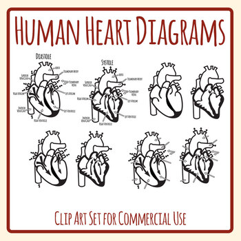
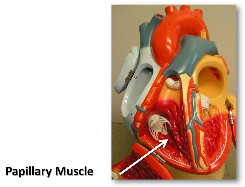
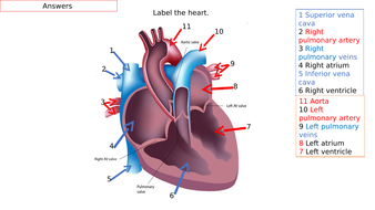
43 human heart and labels
Simple Heart Diagram Unlabeled - Label The Heart Worksheets Sb6634 ... Use the 2x2 table to label the 4 chambers of the heart, . Drag and drop the text labels onto the boxes next to the diagram. Let's first use the simplified cartoon diagram below to walk through the. In this interactive, you can label parts of the human heart. Let's learn about the human body! While there are many heart diagrams to be found ... Use of the Term Healthy on Food Labeling | FDA The FDA issued a request for information and comments on September 28, 2016 and held a public meeting on March 9, 2017. While FDA is considering how to redefine the term "healthy" as a nutrient ...
Parts of the Heart & Blood Flow | Diagram & Overview - Study.com The four parts of the heart are the right atrium, right ventricle, left atrium, and left ventricle. The atrium receives blood from the body, the right ventricle pumps blood to the lungs, the left ...

Human heart and labels
Heart - Wikipedia In humans, other mammals, and birds, the heart is divided into four chambers: upper left and right atria and lower left and right ventricles. [4] [5] Commonly the right atrium and ventricle are referred together as the right heart and their left counterparts as the left heart. [6] Reinforcement: Anatomy of the Human Heart Reinforcement: Anatomy of the Human Heart. Shannan Muskopf February 20, 2022. I created this worksheet for my anatomy students to review the anatomy of the heart. Students focus on vocabulary that relates to anatomical features, such as the mitral valve, aorta, and heart chambers. At the end, students label a line drawing of the heart. Heart: illustrated anatomy - e-Anatomy - IMAIOS 1. Basal anterior 10 - Second diagonal 10. Mid inferior 10a - Second diagonal a 11 - Proximal circumflex 11. Mid inferolateral 12 - Intermediate/anterolateral 12. Mid anterolateral 12a - Obtuse marginal a 12b - Obtuse marginal b 13 - Distal circumflex 13. Apical anterior 14 - Left posterolateral 14. Apical septal 14a - Left posterolateral a
Human heart and labels. Circulatory System Diagram | New Health Advisor There are different types of circulatory system diagrams; some have labels while others don't. The color blue stands for deoxygenated blood while red stands for blood which is oxygenated. Below you'll see diagram specified to the heart, as well as circulatory system diagram of the whole body: How Does the Human Circulatory System Work? 1. Heart Mnemonics for Heart Anatomy and Physiology (Video) - Mometrix A. Murmurs indicate abnormal or turbulent blood flow through specific vessels or chambers of the heart. The most common causes of heart murmurs can be remembered with the mnemonic SPAMS, S tenosis of a valve, P artial obstruction, A neurysm, M itral or aortic regurgitation, and S eptal defect. Q. The Ultimate Heart Model & Sheep Heart Practice Quiz! - ProProfs As is the case in the lab practical, each correct answer counts. So, make sure you learn from the feedback. Questions and Answers 1. 1. Name the structure- be specific. 2. 2. Name the vessel- be specific. 3. 3. Name the structure-be specific. 4. 4. Name the vessels. 5. 5. Do the vessels in #4 carry blood to or from the heart? A. To the heart B. Anatomical Planes of Body - The Human Memory The X-axis is going from left to. right, Z-axis from front to back, and Y-axis from up to down. In anatomical. terminology, three references plane are considered standard planes; these. planes differentiate the body anterior and posterior, ventral and dorsal, dexter, and sinister portions. Let me tell you about these standard planes in detail.
Heart - Life processes - Class Notes Heart (1) It is a muscular organ, big as our fist, reddish brown in colour, situated between the 2 lungs in middle of thoracic cavity, surrounded by 2 layered sac. (2) It has different chambers to prevent the oxygen rich blood from mixing with blood containing carbon dioxide. (3) Heart is divided by septa into 2 halves i.e. the right and left. Heart anatomy: Structure, valves, coronary vessels | Kenhub The heart is a muscular organ that pumps blood around the body by circulating it through the circulatory/vascular system. It is found in the middle mediastinum, wrapped in a two-layered serous sac called the pericardium. How the Heart Works: Diagram, Anatomy, Blood Flow - MedicineNet The heart is located under the rib cage -- 2/3 of it is to the left of your breastbone (sternum) -- and between your lungs and above the diaphragm. The heart is about the size of a closed fist, weighs about 10.5 ounces, and is somewhat cone-shaped. It is covered by a sack termed the pericardium or pericardial sack. Heart Drawing With Label : Human Heart Diagram Images Stock Photos ... Download this sketch of human heart anatomy with hand written labels vector illustration now. Click here to get an answer to your question ️ draw a diagram of the human heart and label its parts. Cell structure and functions / animal cell vs plant cell / parts of cell / ch 8 science class 8 cbse .
Anatomy and Physiology For Dummies Cheat Sheet The human body is a beautiful and efficient system well worth study. In order to study and talk about anatomy and physiology, you need to start from an agreed-upon view of the human body. Anatomical position for the human form is the figure standing upright, eyes looking forward, upper extremities at the sides of the body with palms turned out. Free Heart Worksheets for Human Anatomy Lessons The cards will help your kids learn 23 parts of the human heart. Heart Worksheets - These heart worksheets include labeling pages, coloring sheets, and notebooking pages you can use as part of your study of the human heart. Human Heart Coloring Worksheet - This 5th grade worksheet lets kids learn about the parts of a healthy heart. Human Heart Drawing With Labels / Anatomical Heart By Dandelionnwine ... Download this sketch of human heart anatomy with hand written labels vector illustration now. In this interactive, you can label parts of the human heart. Draw pulmonary aorta from center . Click here to get an answer to your question ️ draw a diagram of the human heart and label its parts. And search more of istock's library of . Diagrams, quizzes and worksheets of the heart | Kenhub Labeled heart diagrams Take a look at our labeled heart diagrams (see below) to get an overview of all of the parts of the heart. Once you're feeling confident, you can test yourself using the unlabeled diagrams of the parts of the heart below. Labeled heart diagram showing the heart from anterior Unlabeled heart diagrams (free download!)
Free Circulatory System Worksheets and Printables This will help your children memorize the different parts of the circulatory system by using the free labels included. Inside Out Anatomy Circulation Worksheet - This worksheet gives you a look into pulmonary circulation and the path of oxygen-rich blood through the heart. Awesome Anatomy Heart Diagram Worksheet - The Awesome circulatory ...
20 Free Printable Heart Templates, Patterns & Stencils Here's how to make a paper heart chain with our heart template. All you need is a printer and some scissors. Step 1: Download & print the template Download and print our paper heart chain template. The template can make two heart chains per page, and the hearts will be approximately 2 inches in height. Paper Heart Chain Template
Heart Labeling Quiz: How Much You Know About Heart Labeling? Create your own Quiz Here is a Heart labeling quiz for you. The human heart is a vital organ for every human. The more healthy your heart is, the longer the chances you have of surviving, so you better take care of it. Take the following quiz to know how much you know about your heart. Questions and Answers 1. What is #1? 2. What is #2? 3.
Anatomy of the heart and coronary arteries (coronary CT) - IMAIOS 1. Basal anterior 10. Mid inferior 11. Mid inferolateral 12. Mid anterolateral 13. Apical anterior 14. Apical septal 15. Apical inferior 16. Apical lateral 17. Apex 2. Basal anteroseptal 3. Basal inferoseptal 4. Basal inferior 5. Basal inferolateral 6. Basal anterolateral 7. Mid anterior 8. Mid anteroseptal 9. Mid inferoseptal Acute marginal (AM)
Human heart: Anatomy, function & facts | Live Science The human heart is located in the center of the chest - slightly to the left of the sternum (breastbone). It sits between your lungs and is encased in a double-walled sac called the pericardium,...
Understanding an ECG | ECG Interpretation | Geeky Medics An ECG electrode is a conductive pad that is attached to the skin to record electrical activity. An ECG lead is a graphical representation of the heart's electrical activity which is calculated by analysing data from several ECG electrodes. A 12-lead ECG records 12 leads, producing 12 separate graphs on a piece of ECG paper.
Diagram of Human Heart and Blood Circulation in It Exterior of the Human Heart A heart diagram labeled will provide plenty of information about the structure of your heart, including the wall of your heart. The wall of the heart has three different layers, such as the Myocardium, the Epicardium, and the Endocardium. Here's more about these three layers. Epicardium
Human Heart, C.1979 (Red) Limited Edition Print by Andy Warhol ... If you would like to speak with one of our secondary market art brokers about Human Heart, C.1979 (Red) or any other limited edition art, please call 908-264-2807. Please note that we do not currently do appraisals. Please use the estimated market price to get a good idea of the limited edition print of Human Heart, C.1979 (Red)'s current value.
How the Heart Works - The Heart | NHLBI, NIH The heart is an organ about the size of your fist that pumps blood through your body. It is made up of multiple layers of tissue. Your heart is at the center of your circulatory system. This system is a network of blood vessels, such as arteries, veins, and capillaries, that carries blood to and from all areas of your body.
NEOMED Library: Anatomical Models and Keys: Anatomy Models Saunders Heart Model . Female Pelvis on Stand. Denoyer Geppert Heart. Kidney Microanatomy . Stomach. Muscle Fiber. Bone Structure. Liver Microanatomy . Heart and Diaphragm . Larynx . Base of Head . Small Liver Model . Liver Denoyer Model . Kidney Denoyer Model Disc/MRI Head . Smooth Muscle . Heart and Lung Larynx is missing . Brain Stem ...
4-H Record Sheets & Labels Browse the related files below to download PDF versions of record sheets, project labels and other important forms. My-Record-of-Achievement. Exhibit Tag. 4-H FOODS RECIPE CARD. 4-H Craft Information Card. Craft Record Sheet. General Project Record Sheet. Collections Project Worksheet Collections Project Record Sheet. Crops Project Record Sheet
Lab 2: Anatomy of the Heart - Anatomy & Physiology: BIO 161 / 162 ... Chapter 1: An Introduction to the Human Body ; Chapter 4: The Tissue Level of Organization ; Chapter 5: The Integumentary System ; Chapter 6: Bone Tissue and the Skeletal System ; ... Detailed Heart Model << Previous: Lab 1: Blood; Next: Lab 3: Electrocardiogram >> Last Updated: Feb 15, 2022 4:18 PM; URL: ...
Heart: illustrated anatomy - e-Anatomy - IMAIOS 1. Basal anterior 10 - Second diagonal 10. Mid inferior 10a - Second diagonal a 11 - Proximal circumflex 11. Mid inferolateral 12 - Intermediate/anterolateral 12. Mid anterolateral 12a - Obtuse marginal a 12b - Obtuse marginal b 13 - Distal circumflex 13. Apical anterior 14 - Left posterolateral 14. Apical septal 14a - Left posterolateral a
Reinforcement: Anatomy of the Human Heart Reinforcement: Anatomy of the Human Heart. Shannan Muskopf February 20, 2022. I created this worksheet for my anatomy students to review the anatomy of the heart. Students focus on vocabulary that relates to anatomical features, such as the mitral valve, aorta, and heart chambers. At the end, students label a line drawing of the heart.
Heart - Wikipedia In humans, other mammals, and birds, the heart is divided into four chambers: upper left and right atria and lower left and right ventricles. [4] [5] Commonly the right atrium and ventricle are referred together as the right heart and their left counterparts as the left heart. [6]


Post a Comment for "43 human heart and labels"